Benefits Of Laser Scanning Microscopy

What Is Confocal Laser Scanning Microscopy
Fluorescence And Confocal Microscopy Ppt Video Online Download
Olympus Fluoview Resource Center Introduction To Confocal Microscopy

2 Schematic Of The Leica Sp2 Tcs Confocal Laser Scanning Microscope 52 Download Scientific Diagram

Confocal Laser Scanning Microscope An Overview Sciencedirect Topics
Confocal Microscopy Confocal Microscope Scanning Systems Olympus Life Science
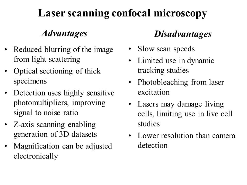
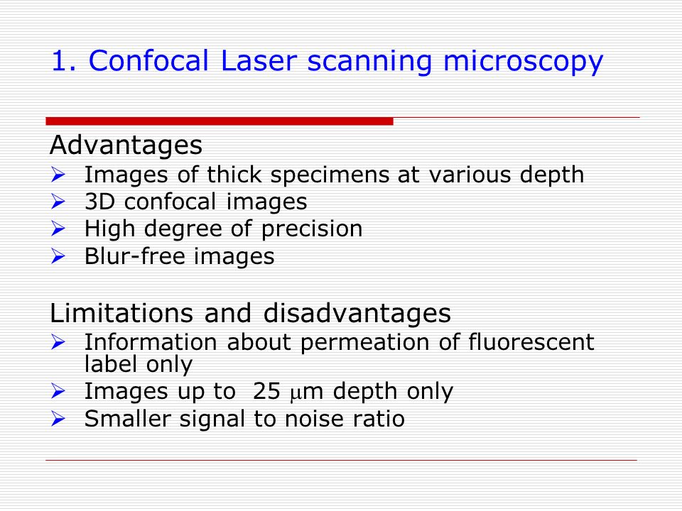
The primary advantage of laser scanning confocal microscopy is the ability to serially produce thin 0 5 to 1 5 micrometer optical sections through fluorescent specimens that have a thickness ranging up to 50 micrometers or more.
Benefits of laser scanning microscopy.
Zeiss Microscopy Online Campus Introduction To Superresolution Microscopy

Zeiss Microscopy Online Campus Introduction To Spinning Disk Microscopy
.png)
Benefits Of Using 3d Laser Confocal Microscopes For Surface Metrology

Overview Of Confocal Laser Scanning Microscopes

Near Field Scanning Optical Microscopy Advantages And Disadvantages

Confocal Microscopy An Overview Sciencedirect Topics

Benefits Of Confocal Microscopy In Modern Life Science Applications Vision Blog
Methods For Monitoring Transdermal Drug Delivery Analytical Radio Nuclear Arn Seminar Jivan Yewle Department Of Chemistry University Of Kentucky Ppt Download
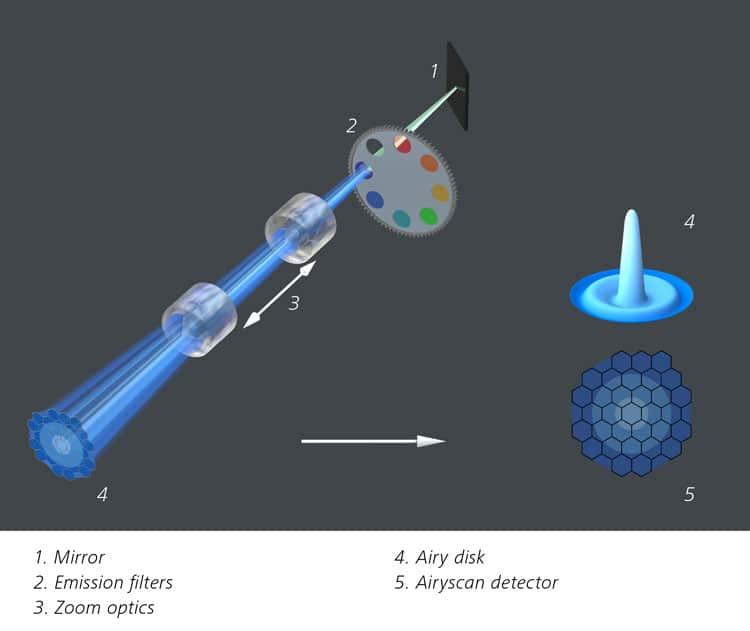
Zeiss Airyscan A Brave New Microscopy World With Sharper Confocal Resolution Bitesize Bio
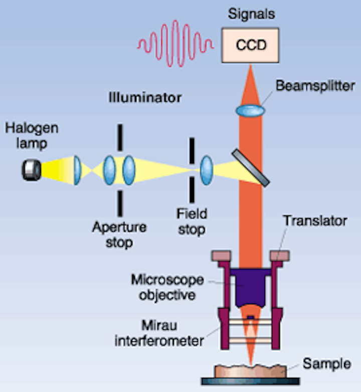
Interferometric Approaches Each Have Advantages Laser Focus World
Pdf Applications Of Confocal Laser Scanning Microscopy Clsm In Foods

Confocal Laser Scanning Microscopy An Overview Sciencedirect Topics

Confocal Microscopy The End Justifies The Means Biocompare The Buyer S Guide For Life Scientists

High Speed 3d Surface Measurement Using Confocal Laser Scanning Microscopy

Confocal Fluorescence Microscopy Lavrentovich Major Reference Works Wiley Online Library
Zeiss Microscopy Online Campus Introduction To Spectral Imaging
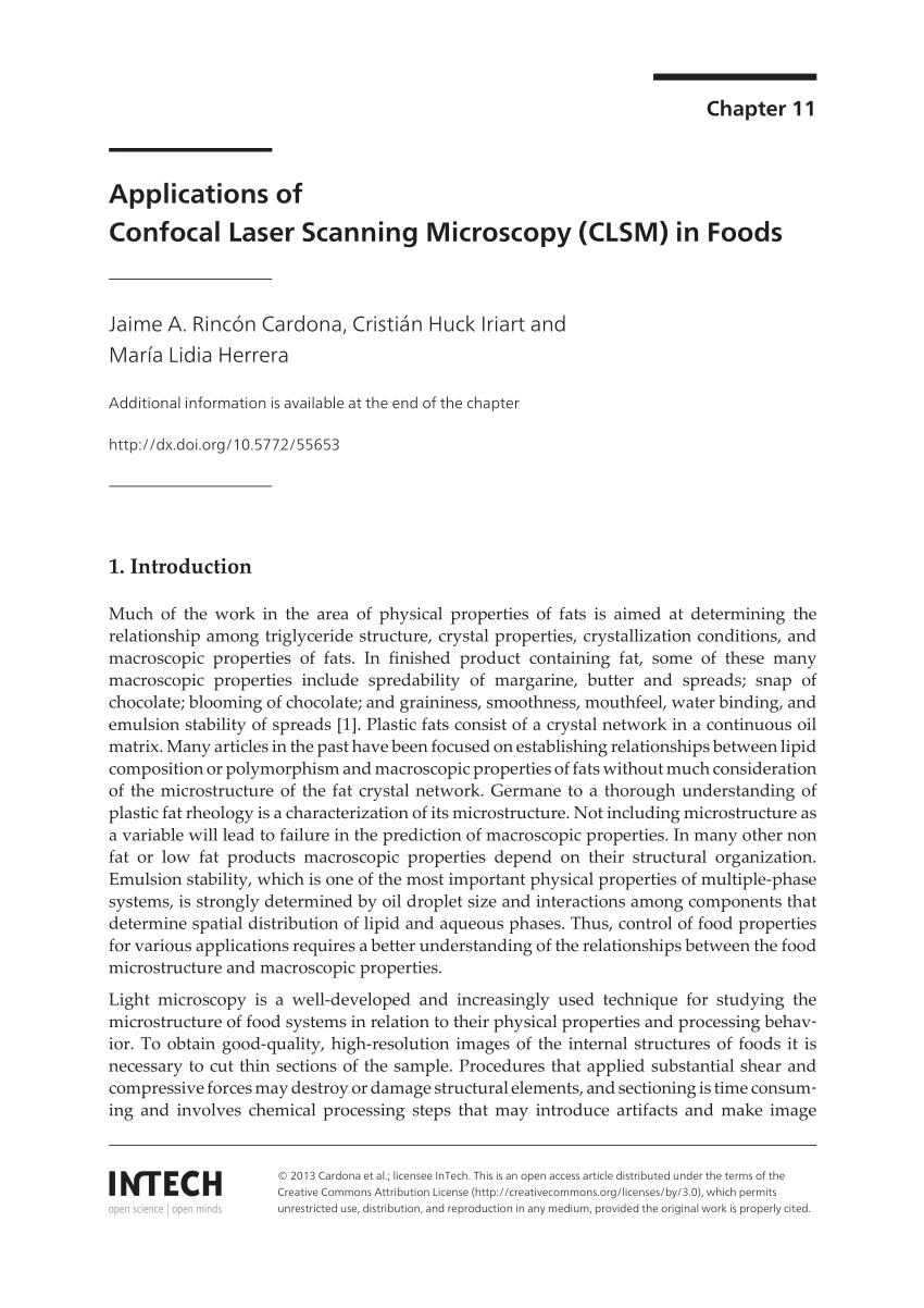
Confocal Microscopy Laser Systems For Confocal Microscopy Olympus Life Science
Spinning Disk Vs Laser Scanning Confocal Microscopes Features Oct 2004 Photonics Spectra
Https Encrypted Tbn0 Gstatic Com Images Q Tbn 3aand9gctgqs1phcsxwxkvi9aampzsqh0nh8wnzi Y6sr 9vm5ljvsbleh Usqp Cau

Recent Advances In Hcs Laser Scanning Cytometry

Confocal Laser Scanning Microscopy An Overview Sciencedirect Topics
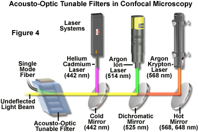
Confocal Microscopy Acousto Optic Tunable Filters Aotfs Olympus Life Science
.jpg)
The Benefits Of Using A Confocal Microscope

Epifluorescence Microscopy An Overview Sciencedirect Topics
Source : pinterest.com
